What is VATS Lobectomy?
Imagine needing a major lung operation but not requiring the long, muscle-dividing incision that patients once associated with chest surgery. That is exactly what Video-Assisted Thoracoscopic Surgery (VATS) lobectomy has made possible. Over the last two decades, VATS has transformed thoracic surgery by allowing surgeons to remove an entire lobe of the lung through a few small incisions using a high-definition camera and specially designed instruments. Today, this minimally invasive technique has become the preferred approach for many patients with early-stage lung cancer, offering the same cancer operation while significantly reducing the physical trauma of surgery. Major international guidelines now recommend minimally invasive surgery over traditional thoracotomy whenever technically feasible for patients with Stage I non-small cell lung cancer. (chestnet.org)
A lobectomy refers to the removal of one of the five lobes of the lungs. The right lung contains three lobes, while the left lung contains two. Removing the affected lobe completely, along with its associated lymph nodes, remains the gold standard treatment for most operable early-stage lung cancers because it provides the best balance between complete tumour removal and long-term survival. Unlike a wedge resection, which removes only a small portion of the lung, or a segmentectomy, which removes a specific anatomical segment, a lobectomy removes the entire diseased lobe while preserving the remaining healthy lung. This approach minimizes the risk of leaving behind microscopic disease and has consistently demonstrated excellent long-term cancer control in appropriately selected patients. (chestnet.org)

For patients, the biggest difference lies in the experience after surgery. Instead of a large incision and significant rib spreading, VATS generally involves two to four small incisions, often between 1 and 4 cm in length. The camera provides a magnified view of delicate structures such as the pulmonary artery, pulmonary veins, bronchus, lymph nodes, and surrounding nerves, enabling meticulous dissection while minimizing tissue injury. Patients frequently experience less postoperative pain, require fewer opioid medications, recover lung function more quickly, and return to daily life sooner than those undergoing conventional open surgery. These advantages have made VATS one of the most important advances in modern thoracic surgery.
Why VATS Lobectomy Has Become the Preferred Treatment for Early Lung Cancer
Only a generation ago, almost every lung cancer operation required a thoracotomy, involving a long incision across the chest with the ribs spread apart to reach the lung. Although thoracotomy remains an essential operation in selected complex cases, advances in imaging, instrumentation, anaesthesia, and surgical expertise have shifted the standard of care toward minimally invasive techniques. Today, VATS lobectomy is considered the preferred surgical approach for most patients with resectable early-stage non-small cell lung cancer because it combines excellent oncological outcomes with a substantially easier postoperative recovery. International guidelines from the American College of Chest Physicians recommend minimally invasive approaches over thoracotomy for clinical Stage I disease whenever appropriate. (chestnet.org)
One of the major concerns when VATS was first introduced was whether smaller incisions might compromise cancer treatment. Surgeons questioned whether lymph node dissection would be adequate, whether tumour handling might be inferior, and whether long-term survival could suffer. Those concerns have now been addressed by extensive clinical research. Recent analyses, including an individual patient data meta-analysis of randomized trials published in The Lancet, demonstrate that VATS provides oncological outcomes equivalent to open lobectomy while preserving its well-recognized benefits of reduced pain, fewer postoperative complications, faster recovery, and improved quality of life. (PubMed)
Patients with reduced lung reserve particularly benefit from VATS. Individuals with chronic obstructive pulmonary disease (COPD), previous smoking-related lung damage, advanced age, or multiple medical conditions often tolerate minimally invasive surgery better because postoperative breathing mechanics recover more rapidly. Smaller incisions facilitate earlier mobilisation, effective coughing, improved physiotherapy participation, and reduced pulmonary complications such as pneumonia. Enhanced Recovery After Surgery (ERAS) pathways now strongly recommend minimally invasive pulmonary resection whenever feasible because these advantages translate into shorter hospital stays and improved patient outcomes. (OUP Academic)
The increasing adoption of VATS also reflects improvements in surgical training and technology. High-definition 4K imaging systems, advanced energy devices, articulating staplers, and sophisticated anaesthetic techniques allow experienced thoracic surgeons to perform increasingly complex resections safely through small incisions. While not every patient is suitable for VATS, and conversion to open surgery remains necessary in selected situations for safety, the vast majority of appropriately selected early-stage lung cancer patients can now undergo definitive treatment through a minimally invasive approach.
Who Needs a VATS Lobectomy?
One of the most common misconceptions is that VATS lobectomy is performed only for lung cancer. Although early-stage non-small cell lung cancer remains the most frequent indication, modern thoracic surgeons use VATS lobectomy for a much broader range of carefully selected conditions. The decision is never based solely on the diagnosis; it also depends on tumour size, location, lymph node involvement, previous infections, overall lung function, and the patient’s ability to tolerate surgery. Every case requires individualized evaluation using high-resolution CT scanning, PET-CT where appropriate, pulmonary function testing, and multidisciplinary discussion.

Lung Cancer
The ideal candidate is a patient with Stage I or selected Stage II non-small cell lung cancer in whom the tumour is confined to a single lobe without extensive invasion of major blood vessels or adjacent organs. These patients often achieve the greatest benefit from minimally invasive surgery because complete cancer removal can be combined with a faster recovery and earlier return to normal life. Even after neoadjuvant therapy, selected patients may still be candidates for VATS in experienced centres.
Benign Lung Diseases
Not every lobectomy is performed for cancer. Certain non-cancerous diseases permanently destroy lung tissue, making surgical removal the best long-term solution. Examples include severe bronchiectasis, destroyed lung following tuberculosis, chronic lung abscesses, localized fungal infections such as aspergilloma, congenital abnormalities including congenital pulmonary airway malformation (CPAM), and symptomatic giant bullae. In these situations, removing the diseased lobe can eliminate recurrent infections, persistent cough, haemoptysis, or chronic breathlessness while preserving the remaining healthy lung.
Metastatic Lung Tumours
Although pulmonary metastases are usually treated with limited resections, carefully selected patients with isolated metastatic disease confined to a single lobe may benefit from a lobectomy. Such decisions are highly individualized and are made after multidisciplinary review involving thoracic surgeons, medical oncologists, radiologists, and other specialists.
It is equally important to recognize that not everyone should undergo VATS lobectomy. Large centrally located tumours invading major vessels, extensive chest wall involvement, bulky mediastinal lymph node disease, dense adhesions from previous infections or surgery, or severe cardiopulmonary compromise may require an open thoracotomy or, in some cases, non-surgical treatment. Choosing the right operation for the right patient remains more important than insisting on a minimally invasive approach. An experienced thoracic surgeon should always prioritize oncological completeness and patient safety over incision size.
Preoperative Evaluation: Preparing for a Safe and Successful VATS Lobectomy
Successful VATS lobectomy begins long before the patient enters the operating theatre. The quality of surgical planning directly influences both safety and long-term outcomes. Every patient undergoes a comprehensive evaluation to answer three critical questions: Can the tumour be completely removed? Can the patient safely tolerate removal of one lung lobe? Is surgery the best treatment compared with other available options? This careful assessment is performed through a multidisciplinary team (MDT) involving thoracic surgeons, pulmonologists, radiologists, medical oncologists, radiation oncologists, pathologists, anaesthesiologists, and specialised thoracic nurses. International guidelines consistently recommend MDT discussion for patients with lung cancer to ensure that treatment decisions are evidence-based and personalised.
The cornerstone of preoperative assessment is a high-resolution contrast-enhanced CT scan of the chest, which provides detailed information about the size, location, and anatomical relationships of the tumour. In most patients with suspected lung cancer, this is complemented by a PET-CT scan, which helps determine whether cancer has spread to lymph nodes or distant organs. PET-CT also assists in identifying unsuspected metastatic disease, preventing unnecessary surgery in patients who would benefit more from systemic treatment. Depending on the tumour’s location, additional investigations such as bronchoscopy, EBUS-TBNA (Endobronchial Ultrasound-Guided Needle Aspiration), CT-guided biopsy, brain MRI, or mediastinoscopy may be recommended to accurately stage the disease before surgery.
Assessing lung function is equally important. Since a lobectomy removes approximately one-fifth to one-third of total lung tissue, surgeons must ensure that the remaining lung can provide adequate respiratory reserve. Patients routinely undergo Pulmonary Function Tests (PFTs), including Forced Expiratory Volume in one second (FEV1) and Diffusing Capacity for Carbon Monoxide (DLCO). When lung function appears borderline, additional tests such as cardiopulmonary exercise testing (CPET), stair-climbing assessment, shuttle walk testing, or quantitative perfusion scanning may be performed. These investigations help estimate predicted postoperative lung function and identify patients who may benefit from prehabilitation before surgery.
Optimising a patient’s overall health is another crucial step. Smoking cessation is strongly encouraged, ideally at least four weeks before surgery, as it reduces postoperative pulmonary complications, improves wound healing, and enhances recovery. Chronic medical conditions such as diabetes, hypertension, heart disease, and COPD are carefully managed in collaboration with the patient’s physicians. Nutritional assessment is increasingly recognised as an important component of surgical preparation because malnutrition and sarcopenia are associated with higher complication rates. Many patients also participate in prehabilitation programmes, combining breathing exercises, physiotherapy, aerobic conditioning, and nutritional optimisation to improve postoperative outcomes. Rather than viewing surgery as a single event, modern thoracic teams treat it as part of a continuum of care that begins weeks before the operation and extends well into recovery.
How is a VATS Lobectomy Performed?
Although patients often imagine lung surgery as a dramatic operation requiring a large incision, the reality of modern VATS lobectomy is remarkably different. The procedure is performed using a high-definition thoracoscopic camera and long, specialised instruments inserted through a few small incisions without routine rib spreading. Despite the smaller incisions, the internal operation remains identical in oncological principles to an open lobectomy. The affected lobe is removed completely along with systematic lymph node dissection, ensuring that cancer surgery remains uncompromised while reducing surgical trauma.
The operation typically lasts between two and four hours, depending on the complexity of the tumour, anatomical variations, previous infections, and whether additional procedures such as chest wall resection or bronchoplasty are required. Throughout the operation, patient safety remains the overriding priority. If unexpected bleeding, dense adhesions, or anatomical difficulties are encountered, conversion to an open thoracotomy is not considered a complication or failure. Rather, it reflects sound surgical judgement aimed at ensuring complete tumour removal while protecting the patient from avoidable risk.
Anaesthesia and Lung Isolation
VATS lobectomy is performed under general anaesthesia with the patient completely asleep and pain-free throughout the procedure. A specialised double-lumen endotracheal tube or, in selected cases, a bronchial blocker is used to isolate the lungs. This allows the anaesthesiologist to ventilate only the healthy lung while temporarily collapsing the lung being operated upon, creating a spacious operative field for the surgeon. Close communication between the anaesthesia and surgical teams is essential because single-lung ventilation influences oxygenation, ventilation pressures, and haemodynamic stability throughout the procedure.
Modern thoracic anaesthesia extends beyond simply administering anaesthetic drugs. Advanced monitoring techniques, lung-protective ventilation strategies, goal-directed fluid therapy, and multimodal pain management all contribute significantly to improved recovery. Many patients receive regional analgesia, such as paravertebral blocks or erector spinae plane blocks, reducing the need for opioid medications after surgery. These techniques enable earlier mobilisation, better breathing, and more effective physiotherapy during the postoperative period.
Patient Positioning and Small Incisions
Once anaesthesia has been established, the patient is positioned on their side with the affected lung facing upward. This lateral decubitus position provides optimal access to the chest cavity while protecting pressure points and maintaining physiological stability. After sterile preparation and draping, the surgeon creates two to four small incisions, depending on the chosen VATS technique and individual patient anatomy.
One incision accommodates the thoracoscope, which projects a magnified, high-definition image of the chest cavity onto large monitors in the operating theatre. Additional incisions allow insertion of specialised instruments designed for delicate dissection around major pulmonary vessels and bronchi. The largest incision, often measuring approximately 3 to 5 centimetres, serves as the utility incision through which the lung lobe is eventually removed using a protective retrieval bag. Unlike traditional thoracotomy, routine rib spreading is avoided, preserving muscles, nerves, and surrounding tissues.
Many experienced thoracic surgeons, including those specialising in advanced minimally invasive surgery, now perform biportal or uniportal VATS in selected patients. These approaches further reduce the number of incisions while maintaining the same oncological principles. The choice depends on tumour characteristics, surgeon experience, and institutional expertise rather than cosmetic considerations alone.
Step-by-Step Removal of the Lung Lobe
After entering the chest, the surgeon performs a systematic exploration to confirm that the disease remains surgically resectable. The lung is inspected carefully for unexpected pleural deposits, additional nodules, or signs of advanced disease that may not have been visible on imaging. Dense adhesions, often resulting from previous infections such as tuberculosis or pneumonia, are gently released to restore access to the pulmonary hilum.
The hilar structures are then dissected in a precise anatomical sequence. Although the order may vary depending on the lobe being removed and surgeon preference, each pulmonary vein, pulmonary artery branch, and lobar bronchus is individually identified, carefully dissected, and divided using advanced endoscopic stapling devices. Maintaining meticulous haemostasis is essential because pulmonary vessels are thin-walled and carry significant blood flow. Once these structures have been divided, the remaining fissure is completed, freeing the affected lobe completely from the rest of the lung.
The excised lobe is placed inside a sterile specimen retrieval bag before being removed through the utility incision. This prevents tumour contact with the wound and facilitates pathological examination. Throughout the operation, the surgical team carefully avoids unnecessary handling of the tumour, adhering to accepted oncological principles designed to minimise the risk of tumour cell dissemination.
Systematic Mediastinal Lymph Node Dissection: A Critical Part of Cancer Surgery
Removing the tumour alone is not sufficient when treating lung cancer. Equally important is the removal and pathological examination of lymph nodes located around the lung and within the mediastinum. This process, known as systematic mediastinal lymph node dissection, is an integral component of every oncologically sound lobectomy and should never be omitted simply because the operation is performed using minimally invasive techniques.
Lymph node dissection serves several essential purposes. First, it allows accurate pathological staging, determining whether microscopic cancer cells have spread beyond the primary tumour. Second, staging information directly influences decisions regarding adjuvant chemotherapy, targeted therapy, or immunotherapy after surgery. Third, complete lymph node removal may reduce the risk of local recurrence in appropriately selected patients. Numerous studies have demonstrated that experienced VATS surgeons can achieve lymph node yields equivalent to those obtained through open thoracotomy, reinforcing the oncological validity of minimally invasive surgery.
The surgeon typically removes lymph nodes from multiple predefined stations according to international staging systems. The exact stations depend on whether the tumour is located in the right or left lung, but comprehensive sampling or complete dissection is essential for optimal staging. Pathologists subsequently examine each lymph node under the microscope, often identifying microscopic metastases that were not visible on preoperative imaging.
Chest Tube Placement and Completion of Surgery
Once the lung lobe and lymph nodes have been removed, the remaining lung is carefully re-expanded under direct vision. The surgeon meticulously checks every staple line and dissection area for bleeding or air leaks. Minor air leaks are relatively common immediately after lung surgery and often resolve spontaneously within the first few postoperative days. If necessary, additional sutures, sealants, or reinforcing techniques may be used to minimise prolonged air leakage.
A chest drain (intercostal tube) is then inserted through one of the existing incisions. Its purpose is to remove air and fluid from the pleural cavity while allowing the remaining lung to fully expand. Before closing the incisions, the anaesthesiologist performs a recruitment manoeuvre to inflate the lung completely, enabling the surgical team to confirm satisfactory expansion.
The incisions are closed with absorbable sutures beneath the skin, leaving only small scars that typically fade over time. Patients are then transferred to the recovery area, where specialised thoracic nurses, physiotherapists, and anaesthesiologists begin the next equally important phase of treatment—enhanced postoperative recovery.
Recovery After VATS Lobectomy: What Patients Can Expect
One of the greatest advantages of VATS lobectomy is that recovery begins much sooner than many patients anticipate. While a lobectomy is undeniably a major operation, the minimally invasive nature of VATS significantly reduces the trauma to the chest wall. Instead of spending days confined to bed, most patients are encouraged to sit up, stand, and even take their first few steps on the day of surgery or the morning after. Early mobilisation is not simply about getting patients moving—it is an evidence-based strategy that reduces the risk of pneumonia, blood clots, muscle weakness, and delayed recovery.

Modern thoracic surgery follows Enhanced Recovery After Surgery (ERAS) principles, a multidisciplinary approach designed to optimise every stage of the patient’s journey. ERAS protocols focus on reducing surgical stress, controlling pain effectively, encouraging early nutrition, and restoring normal function as quickly as possible. Numerous studies have shown that ERAS pathways shorten hospital stays, reduce postoperative complications, and improve patient satisfaction without compromising safety. Rather than relying on prolonged bed rest, today’s approach recognises that controlled activity is one of the most effective forms of postoperative therapy.

Patients are often surprised by how quickly breathing improves once they begin walking regularly and performing breathing exercises. Physiotherapists work closely with patients to teach coughing techniques, deep breathing exercises, and the use of incentive spirometry. These simple interventions help re-expand the remaining lung, clear secretions, and minimise the risk of postoperative atelectasis or chest infections. Recovery is a gradual process rather than a single milestone, and understanding what to expect at each stage helps reduce anxiety and promotes confidence during the healing journey.
The First 24 Hours After Surgery
The immediate postoperative period is carefully monitored by a dedicated thoracic surgical team. After leaving the operating theatre, patients spend a few hours in the recovery unit before returning to the thoracic ward or, in selected high-risk cases, the intensive care unit. Continuous monitoring of oxygen levels, heart rate, blood pressure, and respiratory function ensures that any early issues are identified promptly.
Pain control begins immediately after surgery using a multimodal analgesia strategy. Instead of relying solely on opioids, modern pain management combines several medications and regional anaesthetic techniques. Depending on the patient’s needs, this may include paravertebral blocks, erector spinae plane blocks, local anaesthetic infiltration, paracetamol, non-steroidal anti-inflammatory drugs (when appropriate), and limited opioid use. Better pain control enables patients to breathe deeply, cough effectively, and mobilise earlier, all of which contribute directly to a smoother recovery.
Within hours of surgery, patients are encouraged to begin drinking fluids and gradually resume eating as tolerated. Early nutrition supports wound healing, maintains muscle strength, and reduces postoperative fatigue. Contrary to traditional surgical practices, prolonged fasting after thoracic surgery is no longer recommended unless there is a specific medical reason.
Managing the Chest Drain
Many patients are understandably anxious about the chest drain, often imagining it to be painful or difficult to manage. In reality, the drain is a temporary but essential part of recovery after lung surgery. Its purpose is to remove air and fluid from the pleural cavity while allowing the remaining lung to expand fully. Without adequate drainage, air or fluid could accumulate around the lung, delaying recovery and increasing the risk of complications.
The amount of drainage and the presence of any air leak are monitored several times each day. Advances in digital chest drainage systems now allow surgeons to objectively assess air leaks rather than relying solely on visual observations. These systems can help determine the optimal timing for chest tube removal and may contribute to earlier discharge in selected patients.
For many uncomplicated VATS lobectomies, the chest tube can be removed within 24 to 72 hours, although the exact timing depends on the individual patient’s recovery. Patients usually describe chest tube removal as uncomfortable rather than painful, and the procedure takes only a few seconds. Once the drain has been removed and a chest X-ray confirms satisfactory lung expansion, patients often feel a significant improvement in comfort and mobility.
Pain After VATS Lobectomy
One of the most common questions patients ask is, “How painful is VATS lobectomy?” The honest answer is that some discomfort is expected after any lung operation, but VATS generally causes substantially less pain than traditional open thoracotomy because the ribs are not routinely spread apart. Most patients describe the pain as soreness around the incision sites and a sensation of tightness within the chest rather than severe or unbearable pain.
Pain usually follows a predictable pattern. The first few postoperative days are when discomfort is greatest, after which it gradually improves over several weeks. By six to eight weeks, many patients require little or no regular pain medication. Some patients may experience temporary numbness, tingling, or altered sensation around the incision sites due to irritation of the intercostal nerves. These symptoms generally improve with time, although mild sensory changes can occasionally persist.
Effective pain management is about more than patient comfort. If pain prevents deep breathing or coughing, the risk of pneumonia, atelectasis, and prolonged recovery increases. This is why thoracic surgeons emphasise regular pain medication rather than waiting until pain becomes severe. Good pain control allows patients to breathe normally, participate in physiotherapy, sleep better, and return to daily activities sooner.
When Can You Walk, Climb Stairs and Return to Normal Activities?
Early movement is one of the strongest predictors of successful recovery. Patients are encouraged to walk short distances on the first postoperative day, gradually increasing both the distance and frequency each day. Walking improves circulation, strengthens breathing muscles, reduces fatigue, and helps restore confidence. It is entirely normal to feel breathless initially, but this usually improves steadily as the remaining lung adapts.
Climbing stairs is generally permitted before hospital discharge, provided the patient feels stable and oxygen levels remain satisfactory. Most patients can comfortably manage stairs at home within the first week after surgery. Gentle household activities can usually be resumed within one to two weeks, although heavy lifting should be avoided for approximately six weeks to allow the chest wall to heal properly.
Driving is usually considered safe after three to four weeks, once patients are no longer taking strong opioid painkillers and can comfortably perform an emergency stop without significant pain. Returning to work depends largely on the nature of the occupation. Patients with desk-based jobs often resume work within four to six weeks, whereas those performing physically demanding work may require eight to twelve weeks or longer. Recovery should always be guided by functional ability rather than arbitrary timelines.
Breathing Exercises and Pulmonary Rehabilitation
Recovery from lung surgery is not simply about healing the incisions—it is about helping the remaining lung function as efficiently as possible. Pulmonary rehabilitation plays a vital role in this process. Patients are taught breathing exercises before surgery and continue them throughout the postoperative period. These exercises improve lung expansion, strengthen respiratory muscles, and reduce the likelihood of complications.
An incentive spirometer is one of the simplest yet most valuable tools used after VATS lobectomy. Patients are encouraged to perform slow, deep breaths into the device several times each hour while awake. Although repetitive, this exercise helps prevent collapse of small airways and promotes full expansion of the remaining lung tissue.
For patients with COPD, previous tuberculosis, bronchiectasis, or reduced lung function, formal pulmonary rehabilitation programmes can provide additional benefits. These programmes combine supervised exercise, breathing techniques, education, nutritional counselling, and psychological support. Studies consistently demonstrate that pulmonary rehabilitation improves exercise capacity, reduces breathlessness, and enhances quality of life after lung surgery.
Nutrition After Lung Surgery
Good nutrition is often overlooked, yet it is one of the most important factors influencing recovery. Surgery places the body in a highly metabolic state, increasing the demand for calories and protein. Patients who eat well generally experience better wound healing, maintain muscle strength, and recover more quickly than those with poor nutritional intake.
A balanced diet rich in lean protein, fresh fruits, vegetables, whole grains, and healthy fats supports tissue repair and immune function. Adequate hydration is equally important, particularly during the first few weeks after surgery. Unless restricted for medical reasons, patients should aim to drink sufficient fluids to maintain hydration and help thin respiratory secretions, making coughing easier.
Some patients notice a temporary reduction in appetite during the first week after surgery. Eating smaller, more frequent meals often feels more comfortable than attempting three large meals each day. Nutritional supplements may be recommended for patients experiencing significant weight loss or those with pre-existing malnutrition.
Recovery Timeline After VATS Lobectomy
The following timeline represents a typical recovery for patients undergoing an uncomplicated VATS lobectomy. Individual recovery varies depending on age, lung function, associated medical conditions, and the complexity of the operation.
| Time After Surgery | Typical Recovery Milestones |
| Day 0 | Sitting out of bed, pain control begins, fluids started |
| Day 1 | Walking with assistance, breathing exercises, physiotherapy |
| Days 2–3 | Chest drain removed in many patients, increasing mobility |
| Days 3–5 | Discharge home in uncomplicated cases |
| Week 2 | Walking independently, improving energy levels |
| Weeks 4–6 | Driving, office work, light exercise |
| Weeks 6–12 | Return to most normal daily activities |
| 3–6 Months | Lung function continues to improve, stamina gradually returns |
It is important to recognise that recovery extends beyond the skin incisions. Internal healing continues for several months, and many patients notice ongoing improvements in stamina, breathing, and exercise tolerance well into the first postoperative year.
Expert Perspective from Dr. Kamran Ali
“A successful VATS lobectomy is measured not only by complete tumour removal but also by how quickly and safely a patient returns to normal life. Our goal is to combine oncological excellence with enhanced recovery, allowing patients to regain confidence, independence, and quality of life as early as possible.”
— Dr. Kamran Ali, Associate Director – Thoracic Surgery, Max Super Speciality Hospital, Saket, New Delhi
Benefits of VATS Lobectomy
The transition from open thoracotomy to Video-Assisted Thoracoscopic Surgery (VATS) represents one of the most significant advances in modern thoracic surgery. While the primary objective of a lobectomy remains unchanged—complete removal of the diseased lung lobe with appropriate lymph node dissection—the method by which surgeons achieve this has evolved dramatically. For suitable patients, VATS offers the same oncological principles while substantially reducing the trauma associated with surgery. It is important to emphasise, however, that these benefits are realised only when the operation is performed by an experienced thoracic surgeon who can adhere to meticulous surgical technique without compromising cancer clearance.
Perhaps the most noticeable advantage for patients is reduced postoperative pain. Traditional thoracotomy requires a large incision and rib spreading, which can injure chest wall muscles, ribs, and intercostal nerves. VATS avoids routine rib spreading, resulting in less tissue damage and a lower incidence of severe postoperative pain. Better pain control enables patients to breathe deeply, cough effectively, and participate actively in physiotherapy, all of which contribute to faster recovery and fewer respiratory complications.
The benefits extend well beyond comfort. Numerous studies have demonstrated that patients undergoing VATS lobectomy experience shorter hospital stays, lower rates of postoperative pneumonia, reduced blood loss, decreased need for blood transfusions, earlier mobilisation, and quicker return to daily activities. Smaller incisions also provide superior cosmetic outcomes, although this should never be considered the primary reason for choosing a minimally invasive approach. The true value of VATS lies in preserving physiological function while maintaining uncompromised cancer surgery.
Another important advantage is the ability to begin adjuvant treatment, such as chemotherapy or targeted therapy, sooner when required. Patients who recover more quickly are often better positioned to receive additional treatment without unnecessary delays. In lung cancer, timely initiation of postoperative therapy can be an important factor influencing long-term outcomes, particularly in patients with lymph node involvement or other high-risk pathological features.
Why Experience Matters More Than the Size of the Incision
Patients often assume that minimally invasive surgery is automatically the best choice simply because the incisions are smaller. In reality, the quality of the surgeon and the completeness of the cancer operation are far more important than the number or length of the incisions. A perfectly executed open lobectomy is always preferable to a poorly performed minimally invasive procedure.
Experienced thoracic surgeons understand when VATS is appropriate and when patient safety requires conversion to an open thoracotomy. Complex hilar anatomy, calcified lymph nodes, dense adhesions following tuberculosis or previous infections, unexpected bleeding, or tumour invasion into major vessels may make minimally invasive surgery unsafe. Choosing to convert is a mark of sound surgical judgment rather than technical failure. The priority must always remain complete tumour removal, meticulous haemostasis, and patient safety.
Equally important is the surgeon’s ability to perform a systematic mediastinal lymph node dissection. High-quality lymph node dissection is essential for accurate staging and long-term oncological outcomes. Patients should therefore seek treatment from surgeons and centres with dedicated expertise in thoracic oncology rather than focusing solely on the availability of minimally invasive technology.
VATS Lobectomy vs Open Lobectomy
Although open thoracotomy remains an indispensable procedure for many complex thoracic operations, VATS has become the preferred approach for appropriately selected patients with early-stage lung cancer. Understanding the differences helps patients appreciate why minimally invasive surgery has gained widespread acceptance while recognising that both operations remain valuable tools in modern thoracic surgery.
| Feature | VATS Lobectomy | Open Lobectomy (Thoracotomy) |
| Incision | 2–4 small incisions | Large posterolateral chest incision |
| Rib spreading | No routine rib spreading | Yes |
| Postoperative pain | Usually less | Usually greater |
| Blood loss | Generally lower | Often higher |
| Hospital stay | Typically 3–5 days | Usually 5–8 days |
| Recovery | Faster return to normal activities | Longer recovery period |
| Cosmetic result | Smaller scars | Larger scar |
| Lymph node dissection | Equivalent when performed by experienced surgeons | Standard approach |
| Long-term cancer outcomes | Equivalent in appropriately selected patients | Established gold standard |
It is essential to understand that both operations aim to achieve exactly the same oncological result. The difference lies in the surgical access rather than the cancer operation itself. In experienced hands, VATS provides equivalent tumour clearance and lymph node dissection while offering improved postoperative recovery for suitable patients.
There are still situations in which thoracotomy remains the better choice. Tumours invading the chest wall, major blood vessels, vertebral bodies, or mediastinal structures often require open surgery to ensure complete resection. Similarly, patients with dense scarring from previous operations or severe inflammatory diseases may not be ideal candidates for VATS. The best thoracic surgeons are proficient in both techniques and select the operation based on what offers the safest and most effective treatment for each individual patient.

VATS Lobectomy vs Robotic Lobectomy
The rapid expansion of robotic-assisted thoracic surgery (RATS) has introduced another minimally invasive option for lung resection. Patients frequently ask whether robotic surgery is superior to VATS. The answer is more nuanced than many marketing materials suggest.
Both VATS and robotic surgery avoid thoracotomy, reduce postoperative pain, and allow complete anatomical lobectomy with systematic lymph node dissection. Both techniques have demonstrated excellent short-term and long-term outcomes when performed by experienced thoracic surgeons. The primary differences lie in the instruments and visualisation rather than the fundamental surgical principles.
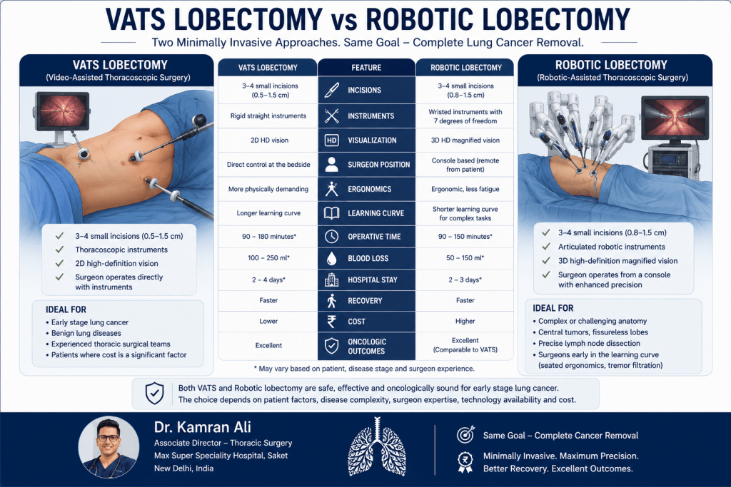
The robotic platform provides the surgeon with a stable three-dimensional magnified view and wristed instruments capable of movements that exceed the range of the human wrist. These features may facilitate precise dissection in confined spaces and can be advantageous in selected complex cases. However, VATS remains an exceptionally effective technique with decades of accumulated evidence supporting its safety and oncological efficacy.
| Feature | VATS Lobectomy | Robotic Lobectomy |
| Camera | High-definition 2D or 3D | High-definition 3D |
| Instrument movement | Straight thoracoscopic instruments | Wristed robotic instruments |
| Surgeon position | Beside the patient | At robotic console |
| Clinical outcomes | Excellent | Excellent |
| Hospital stay | Similar | Similar |
| Pain | Similar | Similar |
| Long-term survival | Comparable | Comparable |
| Cost | Generally lower | Usually higher |
Current evidence suggests that the surgeon’s expertise has a far greater impact on outcomes than the choice between VATS and robotic surgery. A highly experienced VATS surgeon will consistently achieve better results than an inexperienced robotic surgeon, and vice versa. Patients should therefore prioritise surgeon experience, case volume, multidisciplinary support, and institutional outcomes when selecting where to undergo lung cancer surgery.
Possible Risks and Complications of VATS Lobectomy
Although VATS lobectomy is considered a safe and well-established procedure, it remains a major thoracic operation. Every surgical intervention carries potential risks, and patients should understand these before consenting to treatment. Open discussion of complications is an essential part of informed decision-making and reflects responsible surgical practice.
The most common complication is a prolonged air leak, which occurs when air continues to escape from the remaining lung after surgery. Most air leaks resolve spontaneously within a few days, although some patients require a longer duration of chest drainage. Individuals with severe emphysema or fragile lung tissue are at greater risk of prolonged air leakage.
Other recognised complications include:
- Pneumonia
- Atelectasis (partial collapse of the lung)
- Bleeding requiring transfusion or reoperation
- Cardiac arrhythmias, particularly atrial fibrillation
- Wound infection
- Persistent postoperative pain
- Deep vein thrombosis and pulmonary embolism
- Respiratory failure in high-risk patients
- Rare injury to adjacent structures such as the oesophagus, diaphragm, or recurrent laryngeal nerve
Fortunately, the incidence of major complications has declined significantly with advances in surgical techniques, anaesthesia, perioperative care, and ERAS protocols. Careful patient selection, meticulous surgical technique, and experienced multidisciplinary teams remain the most effective strategies for minimising risk.

When Conversion to Open Surgery Becomes Necessary
One of the most misunderstood aspects of minimally invasive thoracic surgery is conversion to thoracotomy. Some patients mistakenly believe that conversion represents a complication or unsuccessful operation. In reality, conversion is often the safest and most appropriate decision when unexpected findings are encountered.
Reasons for conversion may include:
- Significant bleeding from a pulmonary vessel
- Dense inflammatory adhesions
- Calcified hilar lymph nodes
- Difficult tumour anatomy
- Unexpected tumour extension
- Technical limitations preventing safe dissection
An experienced thoracic surgeon makes this decision promptly rather than persisting with a difficult minimally invasive operation that could compromise patient safety. Conversion simply changes the method of access—it does not change the objective of complete cancer removal.
When VATS Lobectomy May Not Be the Best Option
Although VATS has become the preferred approach for many patients, not every lung tumour should be removed thoracoscopically. Appropriate patient selection is fundamental to achieving excellent outcomes.
VATS may not be suitable for patients with:
- Tumours invading the chest wall or diaphragm
- Extensive involvement of major pulmonary vessels
- Invasion of the heart, aorta, or vertebral bodies
- Bulky mediastinal lymph node disease
- Previous extensive thoracic surgery with severe adhesions
- Certain complex sleeve resections (although experienced centres increasingly perform minimally invasive sleeve procedures)
- Medical conditions making single-lung ventilation unsafe
Every patient should undergo detailed review in a multidisciplinary tumour board before finalising the surgical plan. The decision should always be individualised rather than based on a one-size-fits-all philosophy.
Expert Perspective from Dr. Kamran Ali
“Minimally invasive surgery should never become an end in itself. The best operation is the one that removes the cancer completely, preserves as much healthy lung as possible, and offers the safest recovery for the individual patient. At our centre, the surgical approach is tailored to the disease—not the other way around.”
— Dr. Kamran Ali, Associate Director – Thoracic Surgery, Max Super Speciality Hospital, Saket, New Delhi
Long-Term Outcomes After VATS Lobectomy
For most patients diagnosed with early-stage lung cancer, the most important question is not how the operation is performed—it is whether the surgery offers the best chance of cure. Fortunately, decades of research have shown that VATS lobectomy provides long-term cancer outcomes equivalent to open thoracotomy when performed for appropriately selected patients by experienced thoracic surgeons. This means that patients can benefit from the advantages of minimally invasive surgery without compromising the effectiveness of their cancer treatment.
Long-term success depends on several factors beyond the surgical approach itself. These include the stage of the cancer, tumour biology, lymph node involvement, completeness of tumour removal (R0 resection), molecular characteristics of the tumour, and whether additional treatments such as chemotherapy, immunotherapy, or targeted therapy are required. Surgery remains the cornerstone of treatment for most patients with Stage I and selected Stage II non-small cell lung cancer, but it is increasingly integrated into a multidisciplinary treatment pathway.
Modern pathological assessment after lobectomy provides far more information than simply confirming the diagnosis. Pathologists evaluate tumour size, histological subtype, grade, lymphovascular invasion, visceral pleural invasion, spread through air spaces (STAS), margin status, and lymph node involvement. These findings determine the final pathological stage and help guide postoperative treatment recommendations. Patients should understand that the operation is only one part of comprehensive lung cancer care.
Survival Rates Following VATS Lobectomy
Patients frequently search online for survival statistics, hoping to understand what the future may hold. While survival data can provide useful context, it should never be interpreted as an individual prediction. Every patient’s cancer behaves differently, and outcomes vary according to tumour stage, molecular profile, overall health, smoking history, and response to treatment.
In general, patients with Stage I non-small cell lung cancer who undergo complete surgical resection have excellent long-term outcomes, particularly when the tumour is detected early through CT screening or incidental imaging. Five-year survival rates are significantly higher for Stage I disease than for more advanced cancers, highlighting the importance of early diagnosis. Numerous comparative studies have demonstrated that patients undergoing VATS lobectomy achieve disease-free survival and overall survival comparable to those undergoing open lobectomy.
One of the reasons VATS has become widely accepted is that it allows surgeons to maintain the same oncological principles while reducing the physiological stress of surgery. Better postoperative recovery enables patients to resume normal activity sooner and, when necessary, begin adjuvant therapy without unnecessary delays. As lung cancer treatment continues to evolve—with targeted therapies, immunotherapy, and precision medicine playing an increasingly important role—the ability to integrate surgery into a broader treatment strategy has become even more valuable.
Follow-Up After VATS Lobectomy
Completing surgery does not mean the patient’s journey has ended. Regular follow-up is essential to monitor recovery, detect recurrence at an early stage if it occurs, identify new primary lung cancers, and manage any long-term effects of treatment. Follow-up schedules are individualised but generally follow international recommendations.
During the first two years after surgery, patients are typically reviewed every three to six months, including clinical assessment and CT imaging. After this period, annual surveillance is commonly recommended for several years, particularly in patients who remain at risk of developing a second primary lung cancer because of previous smoking or underlying lung disease. Follow-up appointments also provide an opportunity to address persistent symptoms, optimise lung function, reinforce smoking cessation, and discuss lifestyle modifications that support long-term health.
Patients should promptly report new symptoms such as persistent cough, unexplained weight loss, increasing breathlessness, chest pain, coughing up blood, or unexplained fatigue rather than waiting for the next scheduled appointment. While many postoperative symptoms have benign explanations, early evaluation remains the safest approach.
Life After a Lobectomy: Can You Live Normally with One Less Lung Lobe?
One of the most common concerns among patients is whether removing an entire lung lobe will permanently limit their quality of life. The reassuring answer is that most patients adapt remarkably well after lobectomy. The remaining lung tissue gradually expands to occupy part of the empty space, and the body becomes more efficient at using the available lung capacity. Although the lung does not regenerate, functional adaptation allows many patients to resume an active and fulfilling lifestyle.
During the first few weeks, patients often notice reduced stamina and become breathless more easily during physical activity. This is entirely expected and usually improves steadily with regular walking, breathing exercises, and pulmonary rehabilitation where indicated. Individuals who were physically active before surgery often regain excellent exercise tolerance over the following months. Even patients with mild COPD frequently report that, once recovery is complete, they are able to perform most of the activities they enjoyed before surgery.
Returning to work depends on the type of occupation. Office-based professionals commonly resume work within four to six weeks, whereas individuals performing manual labour may require a longer recovery period. Recreational activities such as swimming, cycling, golf, yoga, and moderate gym training are generally encouraged once healing is complete. Air travel is usually safe after satisfactory postoperative recovery and confirmation that there is no residual pneumothorax, although patients should always discuss travel plans with their surgical team.
Lifestyle Changes That Improve Long-Term Outcomes
Surgery removes the tumour, but patients themselves play a vital role in protecting their long-term health. The single most important lifestyle change is permanent smoking cessation. Continuing to smoke after lung cancer surgery increases the risk of recurrence, second primary lung cancers, cardiovascular disease, chronic respiratory illness, and impaired wound healing. Patients who stop smoking—even after many years of tobacco use—derive meaningful health benefits.
Regular physical activity is another key component of recovery. Walking remains one of the most effective exercises during the first few postoperative months, gradually progressing to cycling, swimming, resistance training, or other activities according to individual fitness levels. Maintaining a healthy body weight, consuming a balanced diet rich in protein and fresh vegetables, controlling diabetes and hypertension, and staying up to date with influenza and pneumococcal vaccinations further support long-term wellbeing.
Equally important is emotional recovery. A diagnosis of lung cancer often creates significant psychological stress for both patients and their families. Feelings of anxiety before surveillance scans, fear of recurrence, sleep disturbances, and emotional fatigue are not uncommon. Seeking support from family members, healthcare professionals, counsellors, or patient support groups can be invaluable during recovery. Comprehensive cancer care extends beyond the operating theatre—it encompasses physical, emotional, and social wellbeing.
Why Choose Dr. Kamran Ali for VATS Lobectomy in India?
Choosing a surgeon for lung cancer treatment is one of the most important decisions a patient will make. While technology continues to evolve, successful outcomes depend fundamentally on experience, judgement, multidisciplinary collaboration, and meticulous surgical technique.
As Associate Director – Thoracic Surgery at Max Super Speciality Hospital, Saket, New Delhi, Dr. Kamran Ali specialises exclusively in thoracic surgery, with expertise spanning:
- VATS Lobectomy
- Uniportal and Biportal VATS
- Robotic Thoracic Surgery
- Complex Lung Cancer Surgery
- Sleeve Lobectomy
- Segmentectomy
- Chest Wall Resection
- Mediastinal Tumour Surgery
- Lung Transplantation
- Advanced surgery for post-tuberculosis lung disease, bronchiectasis, empyema, aspergilloma, and other complex thoracic conditions
His practice is built around evidence-based decision-making, multidisciplinary tumour board discussions, meticulous lymph node dissection, and personalised treatment planning. Every patient receives an individualised assessment to determine whether VATS, robotic surgery, or open thoracotomy offers the safest and most effective treatment. The emphasis is never on performing a minimally invasive operation at all costs, but on selecting the surgical approach that provides the best oncological and functional outcome.
Dr. Ali also believes that excellent surgery extends beyond the operation itself. Comprehensive perioperative care—including prehabilitation, ERAS protocols, modern pain management, pulmonary rehabilitation, and structured postoperative follow-up—forms an integral part of achieving optimal recovery and long-term success.
Key Takeaways
- VATS lobectomy is the preferred surgical approach for most patients with early-stage lung cancer when technically feasible.
- It provides equivalent cancer outcomes to open surgery while offering faster recovery, less pain, and shorter hospital stays.
- Systematic mediastinal lymph node dissection remains essential regardless of the surgical approach.
- Not every patient is a candidate for VATS; treatment should always be individualised.
- Choosing an experienced thoracic surgeon and a dedicated multidisciplinary team is more important than the choice of technology alone.
- Recovery continues for several months, but most patients return to an active and productive life after surgery.
Why Patients Trust Dr. Kamran Ali for VATS Lobectomy
Lung cancer surgery is about far more than removing a tumour. It requires careful judgement, meticulous surgical technique, precise anatomical knowledge, and the ability to tailor treatment to each patient’s unique disease. While modern technologies such as VATS and robotic thoracic surgery have transformed the way lung operations are performed, the technology itself is only a tool. The most important determinant of a successful outcome remains the experience and decision-making of the thoracic surgeon.
Dr. Kamran Ali, Associate Director – Thoracic Surgery at Max Super Speciality Hospital, Saket, New Delhi, has dedicated his practice exclusively to diseases of the chest. His work encompasses the complete spectrum of thoracic surgical care, including early and advanced lung cancer, segmentectomy, VATS lobectomy, robotic thoracic surgery, sleeve resections, mediastinal tumours, chest wall tumours, lung transplantation, and complex benign conditions such as bronchiectasis, post-tuberculosis destroyed lung, aspergilloma, empyema, and hyperhidrosis.
Every patient is evaluated through a multidisciplinary approach involving thoracic surgeons, pulmonologists, radiologists, pathologists, anaesthesiologists, medical oncologists, and radiation oncologists whenever required. Treatment recommendations are guided by international evidence and adapted to the individual rather than applying a standard approach to every patient. Whether the best option is VATS, robotic surgery, open thoracotomy, segmentectomy, or even non-surgical treatment, the priority is always the same—achieving the best long-term outcome while preserving quality of life.
Dr. Ali also places strong emphasis on patient education. An informed patient is better equipped to participate in decision-making and often experiences less anxiety before surgery. Every consultation is designed to help patients understand their diagnosis, available treatment options, expected recovery, possible risks, and long-term outlook. The objective is not simply to perform an operation but to guide patients and their families confidently through every stage of treatment—from diagnosis and surgery to recovery and long-term follow-up.
Conclusion
Over the past two decades, VATS lobectomy has fundamentally changed the landscape of lung cancer surgery. What was once considered an innovative technique has become the preferred surgical approach for many patients with early-stage non-small cell lung cancer, supported by robust scientific evidence and international guidelines. By allowing complete anatomical lobectomy and systematic mediastinal lymph node dissection through small incisions without routine rib spreading, VATS offers the dual advantages of excellent oncological outcomes and faster postoperative recovery.
Patients undergoing VATS lobectomy generally experience less postoperative pain, earlier mobilisation, shorter hospital stays, quicker return to work, and improved overall quality of life compared with traditional thoracotomy. These benefits, however, are achieved without compromising the fundamental principles of cancer surgery. Careful patient selection, meticulous surgical technique, and comprehensive perioperative care remain the cornerstones of successful treatment.
It is equally important to recognise that minimally invasive surgery is not appropriate for every patient. Tumour size, anatomical location, lymph node involvement, previous infections, underlying lung function, and overall health all influence the choice of surgical approach. The best outcomes are achieved when treatment decisions are made within a dedicated multidisciplinary thoracic oncology programme led by experienced specialists.
If you or a loved one has been diagnosed with lung cancer and surgery has been recommended, seeking consultation with a dedicated thoracic surgeon can help clarify the diagnosis, review all available treatment options, and determine whether VATS lobectomy is the most suitable approach. Advances in imaging, anaesthesia, surgical technology, and postoperative care mean that many patients today recover more quickly than ever before while receiving world-class cancer treatment based on internationally accepted standards.
Frequently Asked Questions (FAQs)
1. Is VATS lobectomy the best surgery for lung cancer?
For most patients with Stage I and selected Stage II non-small cell lung cancer, VATS lobectomy is considered the preferred surgical approach when technically feasible. It provides the same cancer clearance as open surgery while offering faster recovery, less postoperative pain, and shorter hospitalisation. The final decision depends on tumour characteristics, lung function, and the surgeon’s assessment.
2. How long does it take to recover from VATS lobectomy?
Most patients remain in hospital for three to five days after an uncomplicated VATS lobectomy. Light daily activities usually resume within two to three weeks, office-based work within four to six weeks, and more strenuous activities over the following two to three months. Recovery varies depending on age, lung function, and overall health.
3. Will I be able to breathe normally after one lung lobe is removed?
Yes. Although lung capacity decreases after a lobectomy, the remaining lung adapts remarkably well. Most patients return to an active lifestyle and experience progressive improvement in exercise tolerance during the months following surgery. Pulmonary rehabilitation and regular exercise further enhance recovery.
4. Is VATS lobectomy safer than open thoracotomy?
Both operations are safe when performed by experienced thoracic surgeons. In appropriately selected patients, VATS lobectomy is associated with lower postoperative pain, reduced blood loss, shorter hospital stay, and faster recovery while maintaining equivalent long-term cancer outcomes.
5. What is the success rate of VATS lobectomy?
The success of surgery depends primarily on complete tumour removal (R0 resection), accurate lymph node staging, and the stage of the cancer rather than the size of the incision. In experienced centres, VATS lobectomy has high success rates and excellent long-term survival for appropriately selected patients with early-stage lung cancer.
Related Articles
- Lung Cancer Surgery
- Robotic Lung Cancer Surgery
- Lung Segmentectomy
- Lung Nodules
- Lung Cancer Symptoms
- Bronchoscopy and EBUS
- PET-CT Scan for Lung Cancer
- Lung Cancer Staging
- Recovery After Lung Surgery
- Lung Cancer Treatment in India
References
- Brunelli A, et al. ERS/ESTS Clinical Practice Guidelines on Fitness for Radical Therapy in Lung Cancer. European Respiratory Journal.
- Batchelor TJP, et al. Long-term outcomes of VATS versus open lobectomy for lung cancer. The Lancet.
- National Comprehensive Cancer Network (NCCN). Clinical Practice Guidelines in Oncology: Non-Small Cell Lung Cancer.
- American College of Chest Physicians (CHEST). Guidelines for Early-Stage Non-Small Cell Lung Cancer.
- European Society of Thoracic Surgeons (ESTS). Guidelines for Intraoperative Lymph Node Staging in Lung Cancer.
- Enhanced Recovery After Surgery (ERAS®) Society. Guidelines for Perioperative Care in Lung Resection Surgery.
- International Association for the Study of Lung Cancer (IASLC). TNM Classification of Lung Cancer (9th Edition).
- American Association for Thoracic Surgery (AATS). Consensus Guidelines for Surgical Management of Lung Cancer.


